Fig. 2
From: Atypical white dot syndrome with choriocapillaris ischemia in a patient with latent tuberculosis

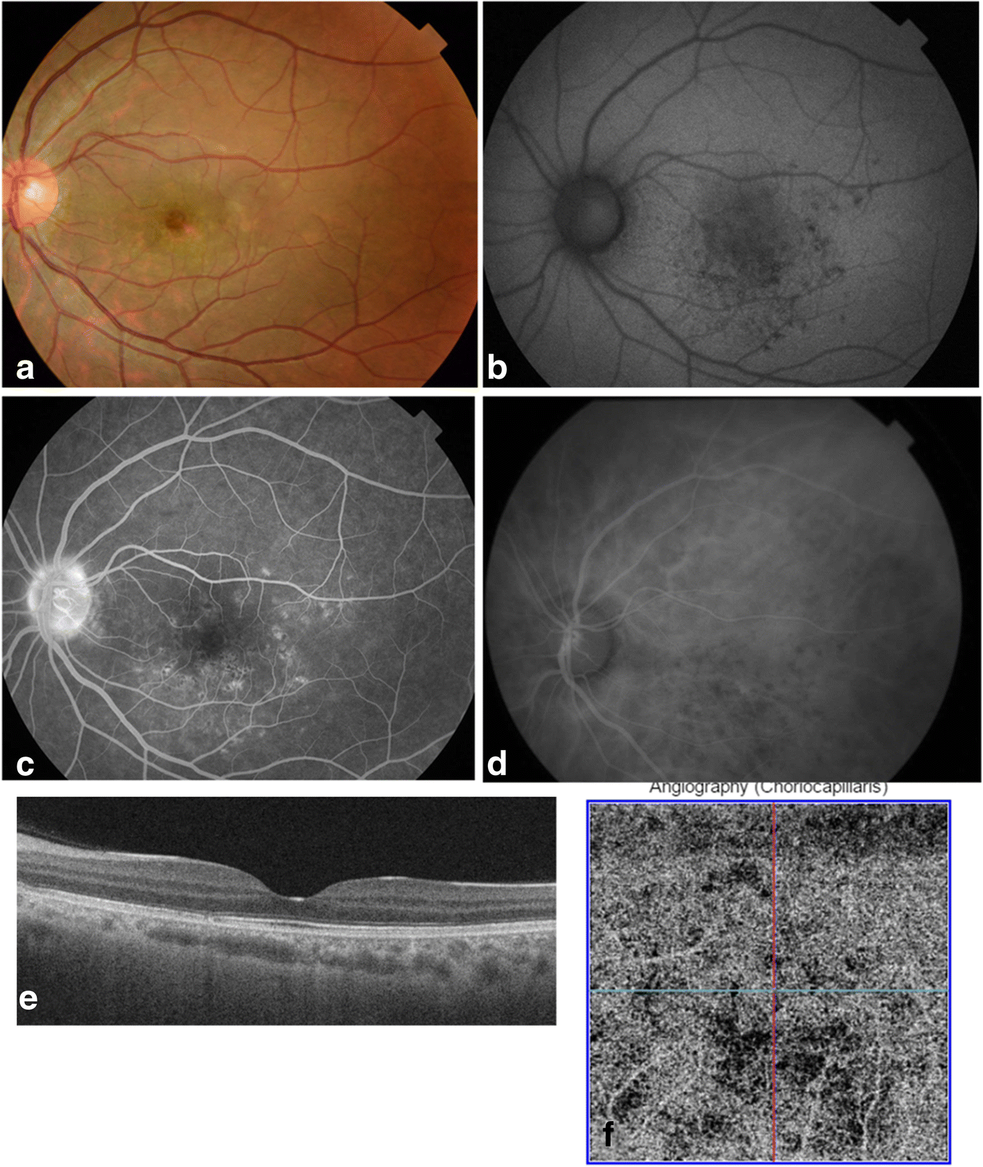
Multimodal imaging findings, 9 months after presentation. a Fundus photograph of the left eye, 9 months after presentation, shows some areas of RPE depigmentation. b Fundus autofluorescence image shows residual punctate areas of hypoautofluorescence. c Late-phase fluorescein angiogram demonstrates residual RPE changes. d Mid-phase indocyanine green angiogram shows small areas of hypofluorescence corresponding to residual scars on fluorescein angiography. e Swept-source OCT image of the left eye, 9 months after presentation, shows resolution of abnormal findings with recovery of a quite normal outer retinal and choroidal aspect (subfoveal choroidal thickness at 200 μm). f Swept-source OCT angiogram of the choriocapillaris reveals markedly improved areas of flow deficit