- Correspondence
- Open access
- Published:
Peripheral retinal cysts in presumed ocular toxocariasis
Journal of Ophthalmic Inflammation and Infection volume 13, Article number: 33 (2023)
Abstract
Typical clinical manifestations of ocular toxocariasis are central posterior granuloma, peripheral granuloma and chronic endophthalmitis. Herein we report the presence of peripheral subretinal cysts in two cases with a presumed ocular toxocariasis (OT).
Photo essay
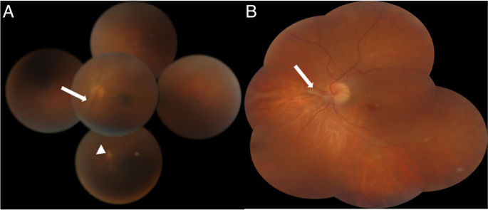
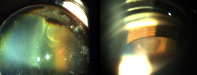
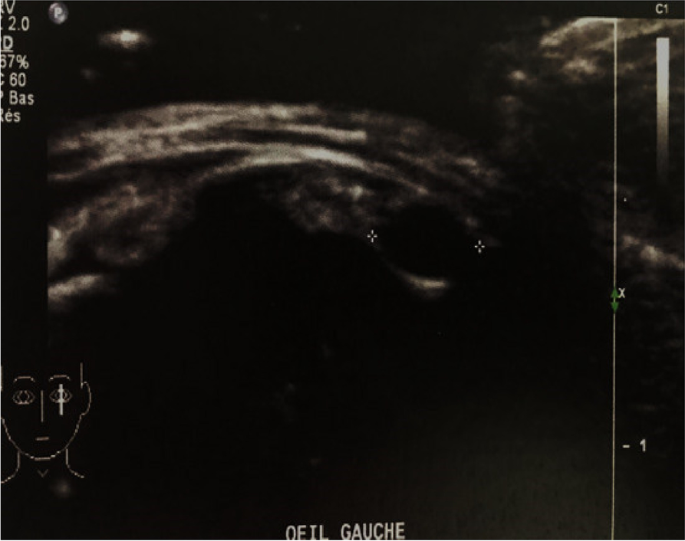
Patients were aged 12 and 20 years old. They were referred to our department for the management of unilateral uveitis. This involved the left eye (OS) for both patients, and the duration of the uveitis before referral was one month for the first patient. The second patient was otherwise asymptomatic. Best corrected visual acuity of the affected left eyes was 20/200 and 20/50, respectively. Anterior segment examination showed the presence of granulomatous anterior uveitis with elevated intraocular pressure, posterior synechiae for the first patient and demonstrated normal results for the second patient. Fundus examination showed the presence of vitritis associated with a vitreoretinal band running from the periphery toward the optic nerve head. A peripheral chorioretinal scar is visible in the first patient. (Fig. 1). Careful examination demonstrated the presence of peripheral retinal cyst in OS for both patients (Fig. 2). Results of ocular examination of the right eye were unremarkable for both patients. Swept source optical coherence tomography showed the presence of macular edema for the first patient. Ultrasound biomicroscopy (UBM) confirmed the presence of peripheral cysts of 2.6 mm and 2 mm in size, respectively, in the second patient (Fig. 3).
Montage color fundus photography in a 12-year-old patient (A) and in a 20-year-old patient (B) with ocular toxocariasis showing a vitreoretinal band running from the periphery toward the optic nerve head (white arrow). A peripheral chorioretinal scar is visible in the first patient (white arrowhead)
Based on clinical presentation, the diagnosis of presumed OT was made and both patients underwent enzyme-linked immunosorbent assay (ELISA) serum test and complete blood count that came back negative with normal eosinophilia respectively for both patients.
Despite the fact that the clinical manifestations of OT are often characteristic, the diagnosis remains challenging, especially in a patient with normal eosinophilia and negative ELISA serology since it’s often repeatedly negative [1].
Peripheral vitreoretinal toxocariasis include vitreal membranes, toxocara granuloma, pseudocysts, thickening of the ciliary body, cystic formation and peripheral retinal detachment, and these lesions were mostly seen and described by UBM, which was considered as an additional diagnostic tool of OT [1, 2]. Pseudocystic formation were otherwise reported to be characteristic of OT [2, 3].
The origin of retinal cysts is not likely to be larva transformation since that during their migration they usually stop growing and end up dying in human tissue [4]. Therefore, some authors suggested that retinal cysts would be caused by a specific OT inflammatory process, often described with chronic uncontrolled intermediate uveitis, leading to vitreous shrinkage and traction resulting in pseudocystic aspect or peripheral retinoschisis [3, 4].
From these two cases, we emphasize the importance of a meticulous peripheral retinal examination in patients with OT in order to look for these peripheral characteristic cysts. In addition, it would be worthwhile to use ultrawide-field imaging and optical coherence tomography, when feasible, in order to ensure better view of the peripheral vitreoretinal changes in OT and provide a more detailed analysis enabling a more accurate diagnosis.
Availability of data and materials
The data used in that case report is available from the corresponding author on reasonable request.
References
Sharkey JA, McKay PS (1993) Ocular toxocariasis in a patient with repeatedly negative ELISA titre to Toxocara canis. Br J Ophthalmol 77(4):253–254 (W Cella, E Ferreira, A M S Torigoe, N Macchiaverni-Filho, V Balarin. Ultrasound biomicroscopy findings in peripheral vitreoretinal toxocariasis. Eur J Ophthalmol. 2004;14(2):132-6)
Tran VT, Lumbroso L, LeHoang P, Herbort CP (1999) Ultrasound biomicroscopy in peripheral retinovitreal toxocariasis. Am J Ophthalmol 127(5):607–609
Despommier D (2003) Toxocariasis: clinical aspects, epidemiology, medical ecology, and molecular aspects. Clin Microbiol Rev 16(2):265–272
Pichi F, Srivastava SK (2017) Nucci Paolo, Baynes K, Neri P, Lowder C Peripheral retinoschisis in intermediate uveitis. Retina 37(11):2167–2174
Funding
No funding was received.
Author information
Authors and Affiliations
Contributions
All the authors contributed to this report: MR, performed the management of the patient. JA and BO reviewed the literature and drafted the manuscript. CM critically reviewed the manuscript and supervised the management. All authors read and approved the final manuscript.
Corresponding author
Ethics declarations
Ethics approval and consent to participate
Not applicable.
Consent for publication
Consent for publication use of images was obtained from the patient.
Competing interests
The authors declare no competing interests.
Additional information
Publisher’s Note
Springer Nature remains neutral with regard to jurisdictional claims in published maps and institutional affiliations.
Rights and permissions
Open Access This article is licensed under a Creative Commons Attribution 4.0 International License, which permits use, sharing, adaptation, distribution and reproduction in any medium or format, as long as you give appropriate credit to the original author(s) and the source, provide a link to the Creative Commons licence, and indicate if changes were made. The images or other third party material in this article are included in the article's Creative Commons licence, unless indicated otherwise in a credit line to the material. If material is not included in the article's Creative Commons licence and your intended use is not permitted by statutory regulation or exceeds the permitted use, you will need to obtain permission directly from the copyright holder. To view a copy of this licence, visit http://creativecommons.org/licenses/by/4.0/.
About this article
Cite this article
Maamouri, R., Jallouli, A., Béizig, O. et al. Peripheral retinal cysts in presumed ocular toxocariasis. J Ophthal Inflamm Infect 13, 33 (2023). https://doi.org/10.1186/s12348-023-00357-y
Received:
Accepted:
Published:
DOI: https://doi.org/10.1186/s12348-023-00357-y


